Learn About Age-related Macular Degeneration

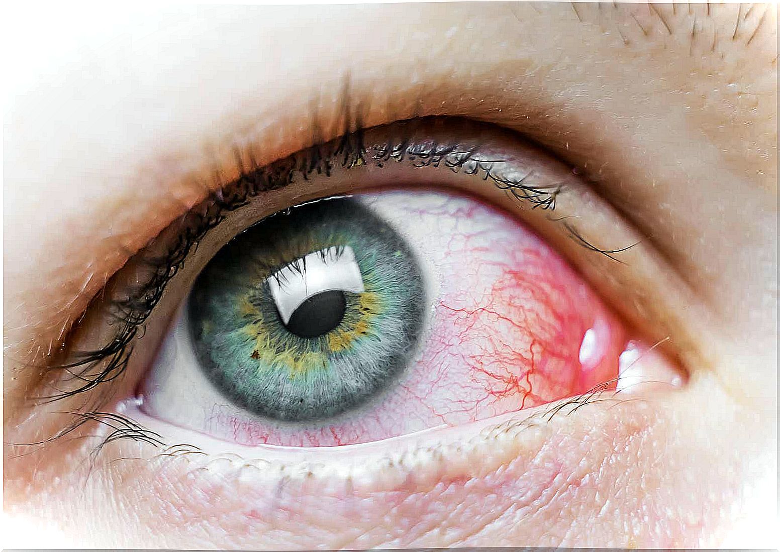
Age-related macular degeneration (AMD) is an eye disorder characterized by the progressive destruction of acute and central vision. This makes it increasingly difficult to visualize the finer details and impedes reading.
This problem is age-related because it is significantly more frequent in people over 50 years of age. Its progress is generally slow, although in some cases it may progress quickly.
Currently, there is great progress being made in the genetic understanding of age-related macular degeneration, and several studies are being carried out to find a solution for this disease, including promising studies related to the transplantation of healthy cells to correct the problem.
What is age-related macular degeneration?
Specifically, age-related macular degeneration is a problem with the retina, which is a light-sensitive tissue and is located deep in the eye. Its function is to convert light and images into electrical impulses that are transmitted to the brain.
The macula is located in the center of the retina. It is a yellow spot that has the function of capturing the most minute details of the vision. Age-related macular degeneration deteriorates central vision but does not affect peripheral vision.
This disease is more common in people with diabetes, overweight, high blood pressure, high cholesterol levels and in people who smoke.
Types of macular degeneration
There are two types of age-related macular degeneration: dry and wet. Wet macular degeneration occurs when blood vessels grow abnormally under the retina. These vessels are fragile and tend to leak blood and fluid.
All of this causes the macula to dislocate from its normal place. Then, damage to the macula is caused, causing it to rapidly degenerate causing loss of vision. The wet type of macular degeneration is the most serious. One of the first symptoms is seeing straight lines as if they were wavy.
Dry macular degeneration is the most common form of this condition. It happens when the cells in the macula, which are sensitive to light, deteriorate. Vision blurs and it becomes more and more difficult to carry out activities that require focused vision. It develops in 3 steps:
- Initial : Initially, the person has no symptoms. When examining the retina, it is possible to see some “druses” or yellow deposits under it.
- Intermediate : here, the patient has many drusen or, in any case, large drusen. It is possible that the vision is already blurred or that more lighting is needed for reading.
- Advanced : in this last phase, there is the presence of many drusen and also deterioration of cells and tissues in the central area of the retina. As a result, the central vision becomes opaque and blurred, making it difficult to read and recognize faces.
Causes of age-related macular degeneration
The exact cause of age-related macular degeneration has not been specified. It is a fact that it is related to the natural deterioration of the organism that occurs over time, but beyond that, there is not much information available at the moment. However, there is a lot of research going on in order to understand more about this subject.
For now, the
- to be caucasian
- smoking
- have blue eyes
- to be female
- have family history
- Functional factors such as a high-fat diet
Diagnosis and prognosis
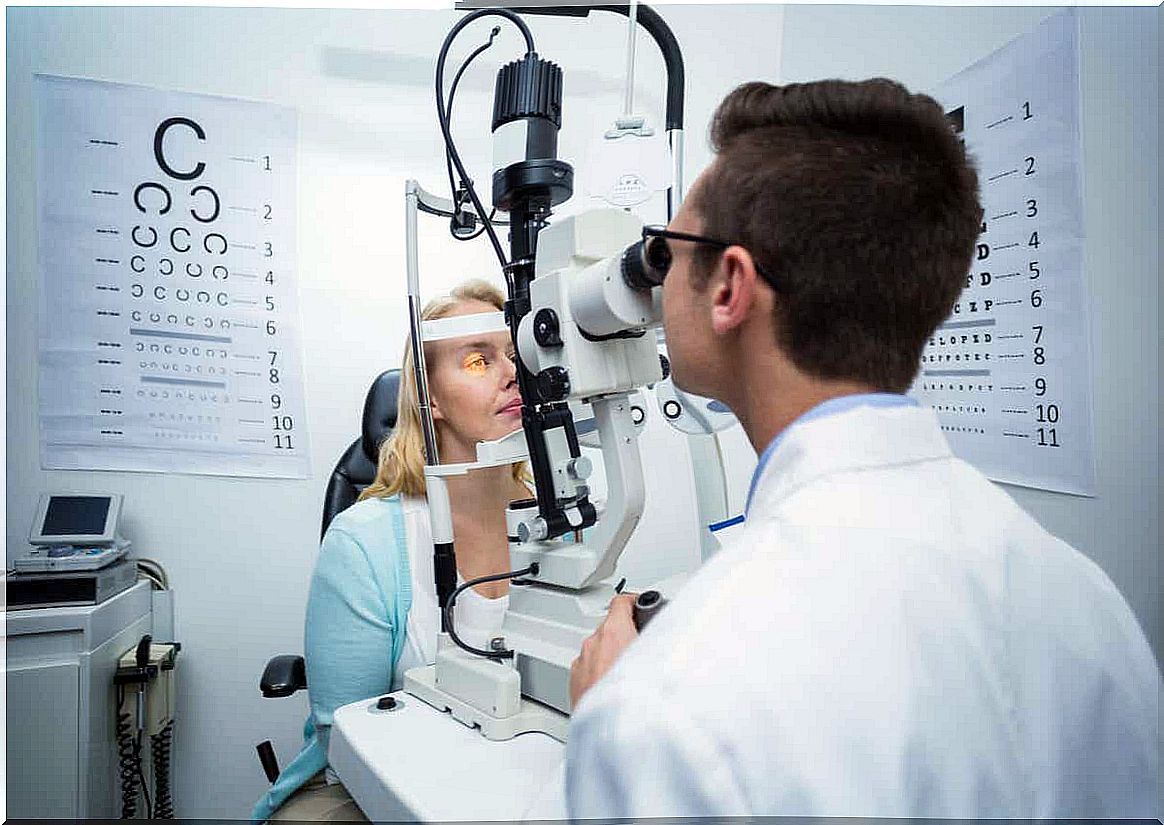
The macular degeneration related to age is detected through an eye exam. Most commonly, this exam includes the following tests:
- Visual acuity test : measures visual ability from different distances.
- Tonometry : measures intraocular pressure with a special device.
- Pupil dilation exam : The doctor examines the retina and optic nerve with a magnifying glass after dropping eye drops on the patient to dilate the pupil.
- Amsler’s canvas : this is a grid similar to a chessboard. The patient performs this examination by covering one eye and looking at a black dot halfway through the grid. Then he repeats the same procedure with the other eye.
The diagnosis of dry macular degeneration is usually positive. However, vision loss in this case is generally not disabling. On the other hand, wet macular degeneration implies a serious loss of vision, making it impossible to carry out activities that require the visualization of small details.
If you notice any of the symptoms mentioned, don’t hesitate to consult a specialist for proper treatment.